Prevention
Modern medicine is increasingly transitioning towards preventive care. This shift towards prevention has also been observed in breast cancer care in recent years, particularly with the discovery of the BRCA gene. Subsequently, multiple genes and risk factors have been identified. Depending on these factors, a personalized screening strategy can be chosen. Therefore, it is crucial to understand these genetic and risk factors.
Diagnosis
I was diagnosed with cancer ... This website serves as a portal designed to assist you and your loved ones in accessing personal information and finding solutions to your concerns.
The primary goal of this website is to offer guidance and support to patients as they navigate their journey toward recovery and improved quality of life. The "Diagnosis" section of our website is divided into two main categories. Firstly, under "Anatomy and Physiology," we provide fundamental knowledge about the breast. Secondly, in the "Tumors and Disorders" section, we delve deeper into various breast-related conditions.
Moreover, we aim to provide information to women who may be concerned about potential breast issues but are hesitant to seek immediate medical advice. Knowledge and information can often offer immediate reassurance if a woman is able to identify the issue herself and determine that no specific treatment is necessary. Conversely, we also strive to educate women who have received a diagnosis of a serious breast condition, such as breast cancer, and wish to approach their doctor well-informed and prepared.
Treatment
The treatment for breast cancer should immediately include a discussion about reconstruction. Our foundation has no greater goal than to raise awareness of this among patients and oncological surgeons. By making an informed decision beforehand, we avoid closing off options for later reconstruction while still considering the oncological aspect. Of course, survival is paramount, and the decision of the oncologic surgeon will always take precedence.
The "Reconstruction or not?" page contains all the information you can expect during an initial consultation before undergoing tumor removal. This page is comprehensive, and your plastic surgeon will only provide information relevant to your situation.
"Removing the tumor" details the surgical procedure itself. This is the most crucial operation because effective tumor removal remains paramount. We guide you through the various methods of removal, a decision often made by a multidisciplinary team comprising oncologists, radiologists, pathologists, radiotherapists, breast nurses, gynecologists, oncological surgeons, and plastic surgeons.
The "Breast Reconstruction" section includes information and illustrations of the different reconstruction options along with corresponding steps.
Revalidation
Those treated for cancer often need a long period to recover.
Cancer is a radical illness with a heavy treatment. Often, people have to deal with psychosocial and/or physical problems afterwards, such as stress, anxiety, extreme fatigue, painful joints, reduced fitness, lymphedema... This can have a major impact on general well-being.
There are rehabilitation programmes offered by most hospitals. We cover some of the major topics here.
Quality of life
Quality of life is a key factor in coping with breast cancer. Therefore, it is important to find coping mechanisms that work, which will be different from patient to patient. For some, it may be finding enjoyment in activities they engaged in prior to diagnosis, taking time for appreciating life and expressing gratitude, volunteering, physical exercise... Of prime importance, studies have shown that accepting the disease as a part of one’s life is a key to effective coping, as well as focusing on mental strength to allow the patient to move on with life. In this section we are addressing some topics that patients experience during and after treatment and we are providing information to address them.
Cancer types
Cancer types
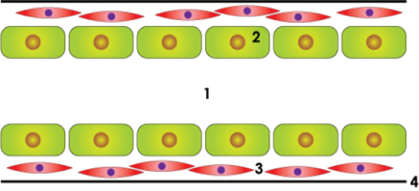
When a breast glandular duct cell transforms into a malignant cell many things need to go wrong. Firstly, the duct cell needs to change its normal behavior and start dividing rapidly, but the cell also needs to survive. Most cells that transform at an abnormally high rate are disrupted by programmed cell death, called apoptosis. This programmed cell death does not occur in cancer cells however.
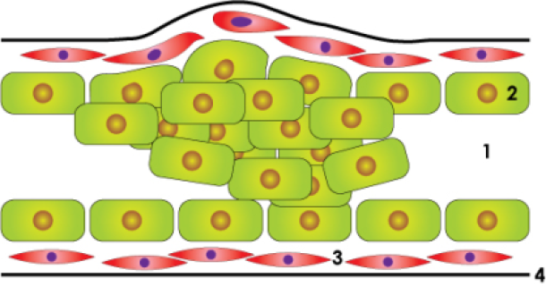
The wild, rapidly dividing cell, starts multiplying. The entire gland tube is filled with malignant cells and the lumen of the tube then disappears. As long as the cancer cells do not break through the wall or basal membrane of the duct, we refer to this cancer as a ‘carcinoma in situ’.
This is a non-invasive tumor with a good prognosis. The fact that the cells do not cross the wall of the basal membrane is crucial. The blood vessels and lymphatic vessels are located in the connective tissue around the gland tubes. As long as the cancer cells do not cross the basal membrane or wall of the gland tube, the cells cannot spread to other organs. The complete removal of a carcinoma in situ therefore guarantees a 100% cure.
Most non-invasive breast cancers do show an increased risk of developing an invasive component. It is therefore imperative that these non-invasive cancers are detected early and treated properly.
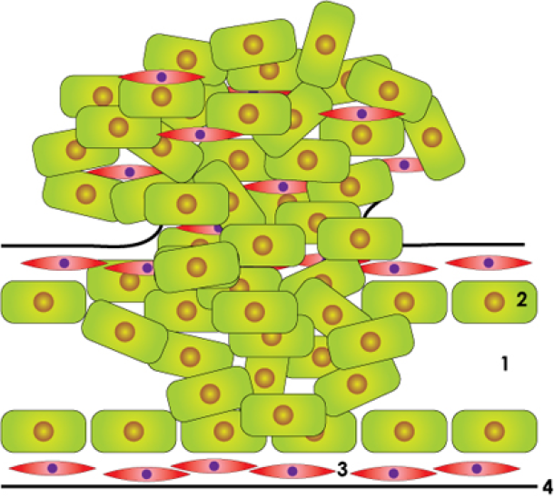
Once the cancer cells migrate through the basal membrane the cancer is called an invasive tumor. From now on the malignant cells can reach the blood and lymph vessels. The cells will form a mass outside the ducts that modifies the normal connective tissue distribution.
On examination of the breast a hard and immobile mass is felt. A malignant tumor is usually not painful. The tumor cells can migrate through the lymphatic vessels to the lymph nodes of the armpit or other areas around the chest. In patients with an invasive tumor, the sentinel node or the lower part of the axillary lymph nodes are removed during surgery and examined for metastases.
Non-invasive breast cancer
Ductal carcinoma in situ (DCIS) and lobular carcinoma in situ (LCIS) are noninvasive breast cancers. They are characterized by a proliferation of malignant cells in the mammary gland and ducts without penetration of the basal membrane.
DCIS is often diagnosed as microcalcification on mammography. DCIS is a precursor of invasive carcinoma and should always be fully removed. There are different degrees of aggressiveness, depending on:
The presence of necrosis
The grade of cell differentiation
The size of the tumor
The presence of microcalcification
LCIS is usually not palpable or visible on mammography. It is often an incidental finding in tissue removed for a different reason. LCIS is usually present in multiple places within the same breast and also both breasts may be affected. If LCIS is present, the risk of developing an invasive cancer is approximately 37%.
Invasive breast cancer
The cells of invasive cancers have the ability to invade and penetrate the surrounding tissues and can produce distant metastases in lymph nodes or other organs. The most frequent types are:
Invasive ductal carcinoma (IDC) (± 75%): This tumor usually presents as a hard mass in the breast and is often surrounded by DCIS.
Invasive lobular carcinoma (ILC) (± 5-10%): This often presents as a poorly defined thickening of the breast and is sometimes difficult to detect, as mammography and ultrasound can not always clearly highlight the tumor. In 90% of patients with ILC, LCIS is also found in the area. ILC is almost always highly hormone-sensitive and has an increased chance of developing in both breasts.
Less frequent types of invasive breast cancer include, medullary carcinoma, mucinous or colloid carcinoma and papillary carcinoma.

normal duct

in situ carcinoma

invasive cancer
Figure above: schematic drawing of a normal lactiferous duct, an in situ carcinoma and an invasive carcinoma: lumen (1), duct cells(2), smooth muscle cells (3), basal membrane (4).
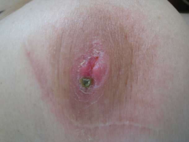
Paget’s disease of the breast is a malignant condition that may have the appearance of eczema of the nipple, with skin changes involving the nipple and sometimes the skin of the breast. Since the condition in itself is often innocuous and limited to a surface appearance, it is sometimes dismissed, although actually indicative of a serious underlying cancerous condition.

Paget's disease of the breast
