Prevention
Modern medicine is increasingly transitioning towards preventive care. This shift towards prevention has also been observed in breast cancer care in recent years, particularly with the discovery of the BRCA gene. Subsequently, multiple genes and risk factors have been identified. Depending on these factors, a personalized screening strategy can be chosen. Therefore, it is crucial to understand these genetic and risk factors.
Diagnosis
I was diagnosed with cancer ... This website serves as a portal designed to assist you and your loved ones in accessing personal information and finding solutions to your concerns.
The primary goal of this website is to offer guidance and support to patients as they navigate their journey toward recovery and improved quality of life. The "Diagnosis" section of our website is divided into two main categories. Firstly, under "Anatomy and Physiology," we provide fundamental knowledge about the breast. Secondly, in the "Tumors and Disorders" section, we delve deeper into various breast-related conditions.
Moreover, we aim to provide information to women who may be concerned about potential breast issues but are hesitant to seek immediate medical advice. Knowledge and information can often offer immediate reassurance if a woman is able to identify the issue herself and determine that no specific treatment is necessary. Conversely, we also strive to educate women who have received a diagnosis of a serious breast condition, such as breast cancer, and wish to approach their doctor well-informed and prepared.
Treatment
The treatment for breast cancer should immediately include a discussion about reconstruction. Our foundation has no greater goal than to raise awareness of this among patients and oncological surgeons. By making an informed decision beforehand, we avoid closing off options for later reconstruction while still considering the oncological aspect. Of course, survival is paramount, and the decision of the oncologic surgeon will always take precedence.
The "Reconstruction or not?" page contains all the information you can expect during an initial consultation before undergoing tumor removal. This page is comprehensive, and your plastic surgeon will only provide information relevant to your situation.
"Removing the tumor" details the surgical procedure itself. This is the most crucial operation because effective tumor removal remains paramount. We guide you through the various methods of removal, a decision often made by a multidisciplinary team comprising oncologists, radiologists, pathologists, radiotherapists, breast nurses, gynecologists, oncological surgeons, and plastic surgeons.
The "Breast Reconstruction" section includes information and illustrations of the different reconstruction options along with corresponding steps.
Revalidation
Those treated for cancer often need a long period to recover.
Cancer is a radical illness with a heavy treatment. Often, people have to deal with psychosocial and/or physical problems afterwards, such as stress, anxiety, extreme fatigue, painful joints, reduced fitness, lymphedema... This can have a major impact on general well-being.
There are rehabilitation programmes offered by most hospitals. We cover some of the major topics here.
Quality of life
Quality of life is a key factor in coping with breast cancer. Therefore, it is important to find coping mechanisms that work, which will be different from patient to patient. For some, it may be finding enjoyment in activities they engaged in prior to diagnosis, taking time for appreciating life and expressing gratitude, volunteering, physical exercise... Of prime importance, studies have shown that accepting the disease as a part of one’s life is a key to effective coping, as well as focusing on mental strength to allow the patient to move on with life. In this section we are addressing some topics that patients experience during and after treatment and we are providing information to address them.
Mastectomy
A mastectomy should be performed when:
The tumor is large compared to the overall size of the breast. When the tumor is very large, chemotherapy can be administered pre-operatively to reduce its size. When the tumor responds to the chemotherapy, breast-conserving surgery may be possible. As a general rule, if more than 1/8 of the breast volume needs to be excised, the aesthetic result after treatment (breast conserving surgery and radiotherapy) will be quite poor, mainly due to the effects of the radiotherapy.
Prophylactic mastectomy (see below): The breast is removed before a cancer occurs. This is recommended for women with a strong genetic predisposition to breast cancer.
Multifocal tumors: when two or more malignant tumors are present in different quadrants of the same breast; or if a malignant tumor is surrounded by satellite lesions; or there is a large area of dysplastic tissue; or there are multiple areas of carcinoma in situ.
Women who have previously been irradiated. This may be the case in patients who have certain forms of leukemia or who have previously undergone breast-conserving surgery combined with radiotherapy and now are confronted with a recurrence.
Severe comorbidities: in elderly women or women who have severe heart or blood vessel disease, severe lung disease or a combination of the above, it is prudent to perform a short procedure. Particularly if the entire breast is removed, one can be sure that no additional surgery will be necessary.
Tumors that have eroded through the skin.
A woman who choses to have her breast completely removed.
In many breast cancer patients, unfortunately, a mastectomy is still required. Fortunately we have moved away from the conventional radical mastectomy and in the majority of cases a modified radical mastectomy is performed.
Even less radical surgery can be offered to patients, depending on their tumor:
Skin sparing mastectomy involves resection of the nipple-areola complex together with the underlying mammary gland but preservation of the skin envelope. Often, the entire gland can be removed through a periareolar incision but if necessary, a short vertical incision can be added in the mid-areolar line.
Areola sparing mastectomy preserves the entire skin envelope and areola but sacrifices the nipple. A vertical incision is made at the mid-areolar line and continued to approximately 1 cm above the infra-mammary fold, to protect this anatomically important structure. The nipple is then removed together with the underlying mammary gland (fig 1.).
In many prophylactic mastectomy cases, a subcutaneous mastectomy is performed, retaining the entire nipple-areola complex. Incisions can be made in the mid-areolar line, in the inframammary fold or through any type of breast reduction/mastopexy pattern incision.
Type of mastectomy or partial gland removal | Anatomical structure that is removed | ||||||
| Breast gland | Nipple | Areola | Breast skin | Axillary lymph nodes | Pectoralis muscle | sternal lymph nodes |
Extended Radical Mastectomy | X | X | X | X | X | X | X |
Radical Mastectomy (Halsted) | X | X | X | X | X | X |
|
Modified Radical Mastectomy (Madden & Patey) | X | X | X | X | X |
|
|
Simple Mastectomy | X | X | X | X |
|
|
|
Skin Sparing Mastectomy | X | X | X |
|
|
|
|
Areola Sparing Mastectomy | X | X |
|
|
|
|
|
Subcutaneous Mastectomy | X |
|
|
|
|
|
|
partial |
|
| +/- |
|
|
| |
small part |
|
|
|
|
|
| |
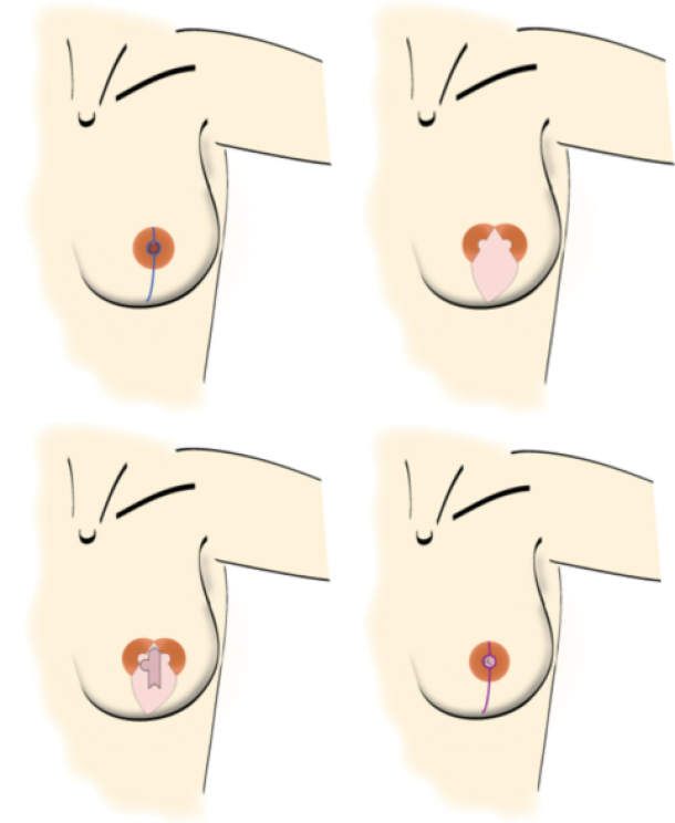
fig.1: Steps in an areola-sparing mastectomy. (a) skin incision for an areola-sparing mastectomy, splitting the areola into two halves and extending to about 1 cm above the inframammary fold; the nipple will be removed. (b) an elliptical skin island of the free flap is fit within the mastectomy skin edges. (c) this skin is used in the second procedure to reconstruct the nipple by means of a modified C-V flap technique. (d) after removal of redundant free flap skin, the wound edges are closed to restore the round shape of the areola and finish with a vertical infra-areolar scar. The new nipple still needs to be pigmented by tattooing during a short third session.
Depending on the patients’ wishes and the breast size and shape, a skin-sparing mastectomy can be combined with either reduction of the skin envelope or a breast lift procedure.
To achieve the best aesthetic result in an immediate reconstruction, it is important to preserve the inframammary fold, the pectoral muscles and the skin envelope during the mastectomy. If any of these anatomical structures are disrupted, they should be repaired before introducing an implant or autologous tissue.
In particular, if there is a deficiency in the skin envelope, then this needs to be addressed. In an immediate autologous reconstruction, the skin can be replaced with skin from the flap. In the case of implant based reconstruction, the extra skin has to be recruited by gradual tissue expansion.
When a patient presents requesting a delayed breast reconstruction, the surgeon has to assess the level of post-ablative damage and the effect of any previous radiotherapy on the chest wall. The more aggressive the initial ablative surgery, the higher the dose of radiotherapy administered (or sensitivity to the radiotherapy), the higher number and type of previous reconstructive attempts and the absence of the nipple-areolar complex, all make the reconstructive procedure more complex and adversely influence the final result.
Examples of different types of mastectomies:

Figure above: schematic drawing and clinical example of a patient who has undergone a modified radical mastectomy without reconstruction.

Figure above: schematic drawing and clinical example of a patient after a skin sparing mastectomy and an immediate autologous breast reconstruction. A nipple reconstruction still needs to be performed.

Figure above: schematic drawing and clinical example of a patient who underwent an areola-sparing mastectomy and an immediate autologous breast reconstruction.

Figure above: schematic drawing and photograph of the same patient as figure 3 after nipple reconstruction and tattoo of the nipple-areolar complex.
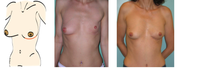
Figure above: schematic drawing and clinical example of a patient who underwent a subcutaneous mastectomy through an infra-mammary incision. She underwent a delayed autologous breast reconstruction through the same incision in a 2nd operative procedure.
References
Radical mastectomy.
ATKINS H. Br J Surg. 1948 Jul;36(141):87-90.
Skin sparing mastectomy: anatomic and technical considerations.
Carlson GW. Am Surg. 1996 Feb;62(2):151-5.
The value of skin-sparing mastectomy.
Kroll SS. Ann Surg Oncol. 1998 Oct-Nov;5(7):660-2.
Twenty-five-year follow-up of a randomized trial comparing radical mastectomy, total mastectomy, and total mastectomy followed by irradiation.
Fisher B, Jeong JH, Anderson S, Bryant J, Fisher ER, Wolmark N. N Engl J Med. 2002 Aug 22;347(8):567-75.
The role of biological markers as predictors of response to preoperative chemotherapy in large primary breast cancer.
Cocquyt VF, Schelfhout VR, Blondeel PN, Depypere HT, Daems KK, Serreyn RF, Praet MM, Van Belle SJ. Med Oncol. 2003;20(3):221-31.
Measuring quality of life in oncologic breast surgery: a systematic review of patient-reported outcome measures.
Chen CM, Cano SJ, Klassen AF, King T, McCarthy C, Cordeiro PG, Morrow M, Pusic AL. Breast J. 2010 Nov-Dec;16(6):587-97.
Immediate conservative breast surgery reconstruction with perforator flaps: new challenges in the era of partial mastectomy reconstruction?
Munhoz AM, Montag E, Arruda E, Brasil JA, Aldrighi JM, Gemperli R, Filassi JR, Ferreira MC. Breast. 2011 Jun;20(3):233-40.
What options for treatment of hereditary breast cancer?
Narod S. Lancet. 2002 Apr 27;359(9316):1451-2.
Conservative mastectomies.
Nava MB, Catanuto G, Pennati A, Garganese G, Spano A. Aesthetic Plast Surg. 2009 Sep;33(5):681-6.
What options for treatment of hereditary breast cancer?
Narod S. Lancet. 2002 Apr 27;359(9316):1451-2.
Elston CW, Ellis IO
Pathological prognostic factors in breast cancer: the value of histological grade in breast cancer. Histopathology (1991) 19:403–410.